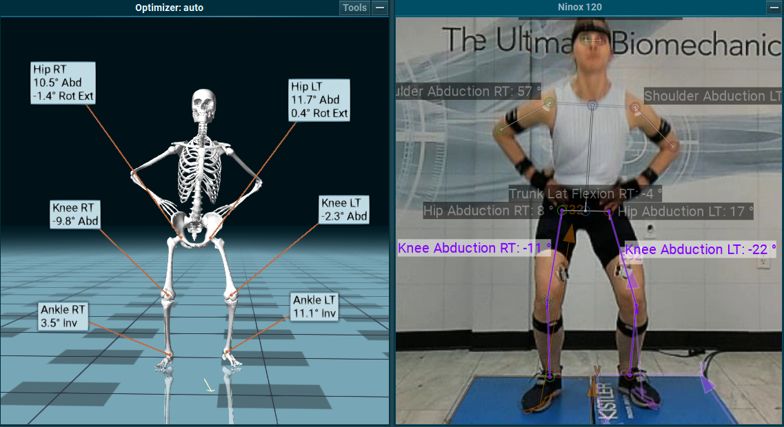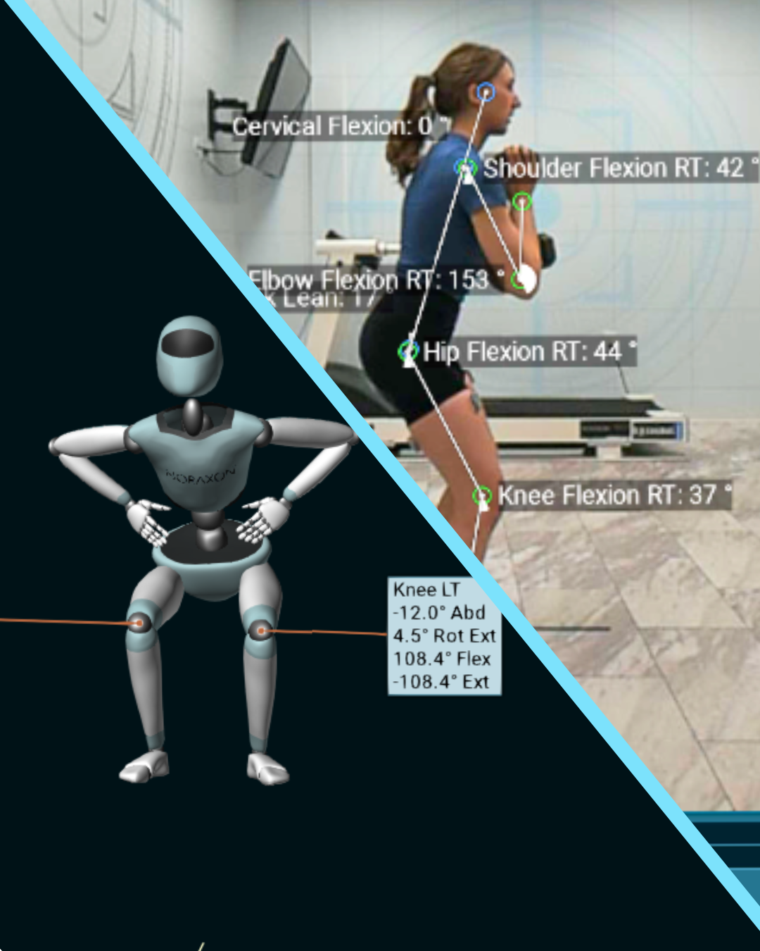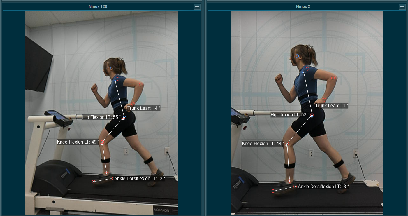
Motion analysis is widely used in clinical, research, and athletic settings to assess joint function, identify movement inefficiencies, and inform rehabilitation or performance programs. While 2D video analysis remains a popular and accessible tool, it comes with important limitations; especially when compared to 3D systems such as IMU-based motion capture.
The Fundamental Difference Between 2D and 3D Systems
- Sagittal or frontal plane only
- Positions and angles are tracked only on visible pixel coordinates
- Lacks depth information and rotational data
- Tracks in all three planes with angular velocity and linear acceleration
- Joint angles using orientation of one segment relative to another
- More anatomically valid representation of joint kinematics


Common Misinterpretations in 2D Analysis
The visual simplicity of 2D analysis is also its greatest flaw. Because it relies on a single camera view, depth and rotation are lost, and joint angles can be severely misrepresented.
Without three-dimensional segmental data, visual judgments are vulnerable to parallax error and misinterpretation.
Practical Considerations for Researchers and Practitioners
Use 2D video analysis when:
- Performing quick movement screens.
- Working in settings with limited equipment.
- You only need gross qualitative insights.
Use 3D IMU analysis when:
- You need to quantify motion across multiple joints and planes.
- You are analyzing rotation (e.g., hip or torso mechanics).
- You are evaluating asymmetries, coupling, or kinetic chains.
When interpreting IMU data:
- Be cautious comparing values directly to what you “see” in video.
- Consider the role of segment rotation in skewing visual interpretation.
- Educate clients and team members about how angles are defined to avoid miscommunication.
In Conclusion
As motion analysis becomes more accessible, it’s critical to understand the capabilities and limitations of the tools we use. While 2D video analysis can offer quick and intuitive feedback, it lacks the anatomical precision needed for accurate kinematic measurement. 3D IMU systems, though more complex, provide segment-relative data that better reflects true joint mechanics.
Visual appearance doesn’t always equal biomechanical reality. By learning to interpret both 2D and 3D data properly, we can make better decisions in clinical, performance, and research contexts.

